At the office of Bella Vida Dental, we invest in diagnostic tools that help our team deliver predictable, efficient care. One of those tools is CBCT (cone-beam computed tomography), a three-dimensional imaging method that reveals anatomy in detail traditional X‑rays can’t show. When used thoughtfully, CBCT enhances diagnosis, treatment planning, and communication between clinician and patient without creating unnecessary complexity.
We rely on CBCT as a focused diagnostic adjunct — not as a substitute for clinical judgment. The images it produces are most valuable when interpreted alongside a full exam, medical history, and your treatment goals. Our approach balances technological capability with patient-centered care so that each recommendation is clear, evidence-based, and tailored to your needs.
Conventional two-dimensional radiographs are excellent for many routine dental needs, but they have limitations when it comes to depth, spatial relationships, and subtle variations in bone and soft-tissue anatomy. CBCT fills those gaps by capturing volumetric data that can be viewed in multiple planes. This three-dimensional perspective helps clinicians detect issues that may be hidden on traditional films, including root morphology, bone defects, and proximity to critical structures.
Because CBCT produces distortion‑free, anatomically accurate views, it improves diagnostic confidence. Clinicians can rotate, crop, and examine thin slices through the jaws to identify patterns that influence treatment decisions. The resulting clarity reduces guesswork and supports more predictable outcomes for complex procedures.
Importantly, CBCT is customizable: field of view, resolution, and exposure settings can be adjusted to match the clinical question. That means we capture only the data needed to answer a specific diagnostic question, which helps limit unnecessary imaging while still delivering rich clinical detail.
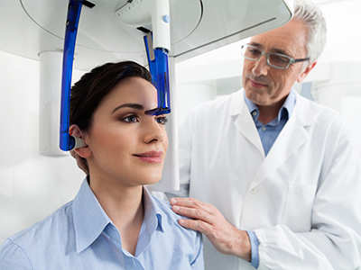
Preparing for a CBCT scan is simple. Patients are usually seated or standing while a cone-shaped X‑ray source and detector rotate around the head. The scan itself takes only a short time—often under a minute for small fields of view—and requires no needles or contrast agents. Because movement can blur details, patients are asked to remain still and follow basic positioning instructions during the brief exposure.
Modern CBCT units are designed to be comfortable and accessible for most patients, including those who have difficulty with traditional panoramic machines. The unit’s open design reduces feelings of confinement, and staff remain nearby to ensure comfort and clear communication throughout the scan.
After acquisition, the volumetric data are reconstructed by specialized software into cross-sectional views and 3D renderings that the dental team can review immediately. This rapid turnaround allows for efficient consultations and clearer discussions about treatment options during the same visit when appropriate.
CBCT is exceptionally valuable for implant planning because it reveals bone volume, density, and the exact relationship of proposed implant sites to nerves and sinus cavities. This level of detail supports precise implant positioning and reduces the risk of complications, contributing to long-term success and function.
Beyond implants, CBCT aids in endodontic assessment by identifying root canal anatomy, fractures, and periapical pathology that may not be visible on two-dimensional films. It also supports oral surgery planning, assessment of impacted teeth, and evaluation of facial trauma where three-dimensional detail changes the surgical approach.
CBCT has growing utility in airway assessment and orthodontic planning as well. By visualizing airway space and craniofacial relationships, clinicians can make better-informed recommendations for patients with breathing-related concerns or complex orthodontic needs. In each application, the scan is used as one component of a comprehensive diagnostic process.
Radiation safety is a top priority when using any X‑ray technology. CBCT units allow clinicians to select the smallest field of view and the lowest acceptable exposure based on the diagnostic task. This ALARA (As Low As Reasonably Achievable) principle guides every imaging decision to keep exposure appropriate and limited.
The effective dose from a focused dental CBCT scan is generally higher than a single periapical X‑ray but substantially lower than most medical CT exams. Our team evaluates the potential benefit of the scan relative to the exposure for each patient and documents the rationale in the clinical record. When a CBCT study will change management or provide information unavailable by other means, the diagnostic value often outweighs the modest increase in exposure.
We also follow strict equipment maintenance and calibration schedules to ensure consistent image quality at the lowest practical dose. Staff are trained in positioning and protocol selection so that every scan is optimized for safety and diagnostic usefulness.
CBCT images are most powerful when integrated into a complete treatment workflow. The three-dimensional data can be used to create surgical guides for implant placement, verify relationships between teeth and surrounding structures, and simulate restorative outcomes. This integration reduces surprises during treatment and allows both clinician and patient to review potential results in advance.
Collaboration is another advantage: CBCT files can be shared with specialists for multidisciplinary cases, enabling more cohesive planning across surgical, orthodontic, and restorative teams. That shared dataset facilitates clearer communication and coordinated care, improving efficiency and patient experience.
At the office level, we emphasize clear explanations and visual aids so patients understand what the images show and how they affect recommended treatment. Engaging patients in the planning process helps set realistic expectations and fosters informed consent grounded in tangible visual information.
CBCT is a sophisticated diagnostic tool that, when used judiciously, elevates the quality of care we provide. If you have questions about whether a CBCT scan is appropriate for your situation, please contact us for more information. We’re happy to discuss how this technology may benefit your treatment plan and overall oral health.


Cone-beam computed tomography (CBCT) is a three-dimensional imaging technique that captures volumetric data of the teeth, jaws and surrounding structures in a single scan. Unlike conventional two-dimensional radiographs, CBCT produces distortion-free cross-sectional views and 3D renderings that reveal depth, spatial relationships and anatomic details not visible on standard films. This added perspective helps clinicians evaluate complex anatomy with greater confidence when needed for diagnosis or treatment planning.
CBCT is not a replacement for routine intraoral X-rays because many everyday diagnostic questions are answered adequately with lower-dose two-dimensional imaging. Instead, CBCT serves as a focused diagnostic adjunct that is chosen when three-dimensional information would change management. Clinicians interpret CBCT findings alongside a clinical exam, medical history and patient goals to form a complete diagnostic picture.
A CBCT scan is recommended when detailed three-dimensional information will affect clinical decision-making, such as implant planning, evaluation of impacted or anomalous teeth, assessment of complex root anatomy and suspected fractures. It is also useful for planning oral surgery, evaluating facial trauma and assessing airway or craniofacial relationships in specialist cases. The decision to obtain a CBCT study is based on the specific clinical question and whether the scan will provide information unavailable from other modalities.
Because CBCT involves higher exposure than a single periapical film, clinicians reserve it for situations where the diagnostic benefit outweighs the modest increase in radiation. Field-of-view and resolution are tailored to the diagnostic task so that only the necessary volume is imaged. This selective approach follows evidence-based guidelines and the ALARA principle to minimize unnecessary exposure.
The scanning process is straightforward and typically takes less than a minute of actual exposure for small fields of view, while setup and positioning may add a few minutes. Patients are asked to remain still while the cone-shaped X-ray source and detector rotate around the head, and staff provide clear instructions and support during the brief acquisition. There are no needles or contrast agents involved, and modern units have open designs to reduce feelings of confinement.
After the scan is acquired, specialized software reconstructs the volumetric data into cross-sectional slices and 3D renderings that the dental team can review immediately. This rapid turnaround often allows the clinician to discuss findings and next steps during the same visit when appropriate. Staff will explain the images in plain language and answer questions about how the scan informs treatment options.
Radiation safety is a core consideration when using CBCT, and clinicians apply the ALARA principle to select the smallest field of view and lowest exposure that will answer the clinical question. A focused dental CBCT scan generally delivers a higher effective dose than a single intraoral film but is substantially lower than most medical CT examinations. Equipment selection, protocol optimization and proper training all contribute to keeping doses as low as reasonably achievable.
Practices document the clinical rationale for each scan and use maintenance and calibration schedules to ensure consistent image quality at minimal dose. Staff training in positioning and protocol selection helps prevent repeat scans caused by motion or incorrect settings. When a CBCT study will change management or reveal information not available by other means, the diagnostic benefit typically justifies the measured exposure.
CBCT provides precise three-dimensional information about bone volume, bone quality and the spatial relationship of proposed implant sites to critical structures such as the inferior alveolar nerve and maxillary sinuses. This level of detail supports accurate implant positioning and depth control, which reduces the risk of complications and helps preserve surrounding anatomy. The ability to visualize bone contours and nearby anatomy preoperatively contributes to more predictable restorative outcomes.
CBCT datasets can be used with planning software to design virtual implant positions and fabricate surgical guides that transfer the plan to the operatory with high fidelity. Using guided surgery reduces intraoperative uncertainty and can streamline implant placement while maintaining safety. When combined with a comprehensive prosthetic plan, three-dimensional imaging helps align surgical and restorative goals for long-term success.
Yes. CBCT can reveal complex root canal anatomy, additional canals, root fractures and periapical pathology that may be missed on two-dimensional films. These three-dimensional views are particularly valuable in retreatment cases, teeth with unusual anatomy and when persistent symptoms are present despite normal-looking 2D radiographs. Identifying anatomy and pathology more clearly helps clinicians plan more effective endodontic approaches.
Interpreting CBCT in endodontics requires attention to resolution and artifact potential, so images are considered alongside clinical findings and pulp testing. High-resolution small-field scans are often selected for targeted endodontic questions to maximize detail while limiting exposure. When necessary, the dentist may consult an endodontic specialist to ensure accurate interpretation and optimal treatment planning.
CBCT has limitations including reduced soft-tissue contrast compared with medical CT, susceptibility to metal artifacts and image degradation from patient movement. It is not the optimal modality for detailed soft-tissue pathology or when contrast-enhanced imaging is required. Additionally, very small lesions or subtle changes may still be better evaluated with complementary radiographic techniques or clinical assessment.
For routine screening or simple diagnostic questions, two-dimensional imaging often provides sufficient information with lower radiation exposure. The clinician weighs diagnostic yield against exposure and selects CBCT only when the expected benefit is clear. This selective approach preserves patient safety and ensures imaging is used judiciously as part of a comprehensive evaluation.
CBCT datasets are stored in standard DICOM format and can be imported into planning software to produce cross-sections, 3D renderings and virtual simulations that inform surgical and restorative workflows. These digital files facilitate the design of surgical guides, prosthetic mock-ups and orthodontic simulations that help align multidisciplinary teams around a single plan. The visual nature of CBCT also improves patient education by allowing clinicians to show specific anatomic findings and discuss options clearly.
When multidisciplinary input is needed, images can be securely shared with oral surgeons, orthodontists, endodontists and laboratories to support coordinated care. This shared dataset reduces ambiguity and enables specialists to provide focused feedback based on the same anatomic information. Clear communication supported by three-dimensional imaging contributes to safer, more efficient treatment pathways.
The practice follows routine equipment maintenance, calibration and quality-control procedures to maintain consistent image performance and optimize dose. Staff are trained in correct patient positioning, protocol selection and artifact reduction techniques so that each scan answers the clinical question with minimal need for repeats. These operational practices help deliver reliable images that support evidence-based decision-making.
Interpretation is performed in the context of a full clinical examination and medical history, and clinicians consult specialists when complex findings require additional expertise. Documenting the diagnostic rationale and integrating CBCT results with other diagnostic data ensures that imaging informs an appropriate, individualized treatment plan. Clear explanations and visible images also help patients understand the role of the scan in their care.
Preparation for a CBCT scan is minimal: patients should remove removable metal such as jewelry, eyeglasses and removable dental appliances that could produce artifacts. Women should inform the clinician if they are pregnant or suspect pregnancy so that the need for imaging can be carefully evaluated and alternatives considered. No dietary restrictions or injections are required for routine dental CBCT scans.
After the scan, the clinician reviews the images with the patient and explains how the findings affect treatment options and next steps. Copies of images or reports can be shared with other providers on request to facilitate continuity of care. If additional imaging or specialist consultation is indicated, the dental team will discuss the rationale and coordinate timely follow-up.

Ready to schedule your next dental appointment or have questions about our services?
Contacting Bella Vida Dental is easy! Our friendly staff is available to assist you with scheduling appointments, answering inquiries about treatment options, and addressing any concerns you may have. Whether you prefer to give us a call, send us an email, or fill out our convenient online contact form, we're here to help. Don't wait to take the first step towards achieving the smile of your dreams – reach out to us today and discover the difference personalized dental care can make.