Digital radiography has become a cornerstone of modern dental care, replacing film-based x-rays with fast, precise digital imaging. This technology uses electronic sensors and computer processing to produce high-resolution images of teeth, roots, and surrounding bone. For patients and clinicians alike, digital radiography means clearer diagnostics, quicker decision-making, and a more comfortable experience during visits to the office.
At its core, digital radiography captures x-ray data using a compact electronic sensor rather than photographic film. The sensor converts x-ray photons into a digital image that can be displayed immediately on a computer screen. This instant access helps clinicians identify cavities, bone loss, impacted teeth, and other conditions with greater confidence than older film methods.
Because digital images are high in contrast and can be enhanced with software, clinicians can adjust brightness, contrast, and magnification to reveal subtle details that may be harder to see on film. These enhancements do not change the underlying data; they simply make diagnostic features easier to recognize without exposing patients to repeat imaging.
For patients, the improved clarity and diagnostic confidence translate into better-targeted treatment plans. Instead of relying on multiple or unclear film images, the clinician can focus on a single, adaptable digital image that supports precise evaluations and explanations during consultations.
Digital sensors are designed to capture x-ray information with consistent sensitivity across the image area, producing uniformly sharp results. Unlike film, whose quality can vary with development processes, digital sensors deliver reproducible outcomes visit after visit. This consistency is especially valuable for monitoring changes over time, such as tracking bone levels or the progress of restorative work.
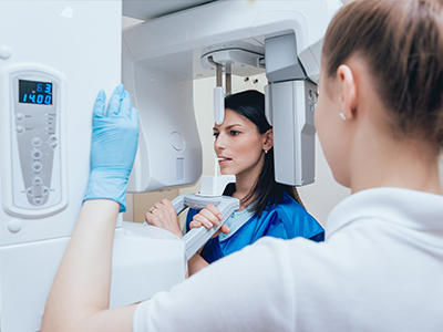
There are two common sensor styles: intraoral panels that sit briefly in the mouth, and small phosphor plates that are scanned after exposure. Both approaches yield images that are readily compatible with practice software. The choice of sensor often depends on patient comfort, clinical needs, and the specific capabilities of the practice’s imaging system.
Clinicians can also use image stitching and panoramic composites to view larger areas when needed, without increasing discomfort for the patient. These integrated imaging options provide comprehensive views that support complex treatment planning while keeping procedures efficient and patient-friendly.
One of the most practical advantages of digital radiography is immediacy. Images appear on-screen within seconds of exposure, allowing clinicians to review results with patients during the same appointment. This immediacy improves the quality of the clinical conversation and helps patients understand findings in real time, using the image as a visual aid.
Once acquired, digital images are stored in the practice’s electronic health record system, where they can be organized, annotated, and retrieved quickly. Secure storage reduces the risk of lost or misfiled films and makes it easier to compare past and present images for long-term monitoring of oral health. Many practices also implement encrypted backups and access controls to protect patient privacy.
Digital files are also convenient for consultations. When coordination with a specialist or a referring provider is needed, images can be shared securely and efficiently, so clinical decisions proceed without unnecessary delays. This streamlined workflow benefits patients who require multidisciplinary care or second opinions.
Digital radiography typically requires less radiation exposure than traditional film x-rays because digital sensors are more efficient at capturing x-ray photons. Lower exposure means increased safety for patients, especially for those who need periodic radiographs as part of ongoing care. Reduced repeat imaging due to clearer initial exposures further minimizes cumulative radiation over time.
For dental teams, the streamlined digital workflow reduces handling of physical materials and exposure to darkroom chemicals historically used in film development. This change improves safety and environmental conditions within the office while simplifying routine procedures for staff.
Practices that embrace digital imaging still follow standard radiographic safety protocols—such as using lead aprons when appropriate, collimation, and modern x-ray units—to keep doses as low as reasonably achievable while obtaining diagnostic-quality images.
Replacing film with digital imaging eliminates the need for chemical developers and paper-based storage, reducing the environmental footprint of radiography. Fewer consumables mean less waste and fewer supplies to manage, which makes clinical operations more sustainable and easier to maintain.
Digital images also support improved care coordination. When images are part of a comprehensive digital record, they can be integrated with other advanced tools—such as intraoral cameras, digital impressions, and 3D scans—to create a unified view of a patient’s oral health. This integration aids in planning restorative, implant, and orthodontic treatments with greater precision.
For patients, the combined effect is smoother visits and clearer communication. Digital records simplify follow-up, allow for more informed second opinions, and support continuity of care across providers, all of which contribute to better outcomes and a more predictable treatment experience.
At Bella Vida Dental, we use digital radiography to support thoughtful, well-documented diagnoses while prioritizing patient comfort and safety. If you’d like to learn more about how digital imaging informs your care, please contact us for more information.


Digital radiography uses compact electronic sensors and computer processing to capture and display x-ray images of teeth, roots, and surrounding bone. The sensor converts x-ray photons into a digital file that appears on a computer screen within seconds, enabling immediate review. This approach replaces film and streamlines diagnostics during the same appointment.
Images are high in contrast and can be enhanced with software tools to reveal subtle details without altering the underlying data. Clinicians can adjust brightness, contrast, and magnification to support more confident evaluations. Patients benefit from clearer explanations and faster decision-making when images are reviewed together with the clinician.
Unlike film x-rays that require chemical processing, digital radiography produces images instantly and eliminates darkroom steps. Sensors provide consistent sensitivity across the image area, avoiding variability tied to film development. The result is reproducible image quality visit after visit for reliable comparisons over time.
Digital files are easy to store and retrieve, reducing the risk of lost or misfiled images and simplifying long-term monitoring. Software enhancements improve diagnostic clarity without additional exposures. The workflow is generally faster and more environmentally friendly than film-based systems.
Digital radiography delivers higher-resolution images and tools that help clinicians detect cavities, bone changes, impacted teeth, and other conditions earlier and more accurately. Being able to enhance and magnify specific areas allows for targeted evaluation of suspicious findings. This precision reduces the need for repeat imaging and supports clearer clinical decisions.
Digital images integrate easily with practice software to support restorative, implant, and orthodontic planning and to track changes over time. At Bella Vida Dental, these images are used to explain findings and to develop individualized treatment strategies that align with each patient’s goals. Secure comparisons between past and current images improve the predictability of care.
There are two common intraoral sensor styles: direct electronic panels that connect to a computer and small reusable phosphor plates that are scanned after exposure. Both capture diagnostic-quality images and are compatible with practice imaging systems. The choice depends on clinical needs, equipment, and the clinician’s preference.
Patient comfort varies with mouth size, gag reflex, and the specific sensor design, so clinicians select the option that minimizes discomfort while delivering the needed image quality. Positioning aids and careful technique further improve comfort and image consistency. Your dental team will discuss the approach that best fits your visit.
Digital sensors are more efficient at capturing x-ray photons, which generally allows for lower radiation exposure compared with traditional film techniques. Modern dental practices follow radiation safety principles and use up-to-date units, collimation, and shielding to keep doses as low as reasonably achievable. Reduced repeat imaging due to clearer initial exposures also lowers cumulative exposure over time.
Staff follow established protocols including the use of lead aprons when appropriate and limiting exposures to clinically necessary images. Patients who are pregnant or have specific medical concerns should inform the team so precautions can be taken. The focus is always on obtaining diagnostic-quality images while protecting patient safety.
After acquisition, digital radiographs are stored in the practice’s electronic health record where they can be organized, annotated, and retrieved quickly. Secure storage reduces the chance of lost films and makes it easier to compare images over time for ongoing monitoring. Many practices use encrypted backups and access controls to protect patient privacy.
When consultation with a specialist or a referring provider is required, images can be shared through secure, HIPAA-compliant channels to support coordinated care. This efficient sharing helps clinical decisions move forward without unnecessary delays. Patients benefit from faster referrals and clearer communication among providers.
High-resolution digital images provide essential information about bone levels, tooth roots, and anatomical landmarks that influence implant and restorative planning. Clear views of the surgical site and surrounding structures help clinicians select appropriate implant sizes and positions. This precision reduces uncertainty and supports safer, more predictable procedures.
Digital radiographs can be combined with CBCT scans, digital impressions, and treatment planning software to create an integrated view of the mouth. That integration aids in designing restorations and guides for implant placement with greater accuracy. The result is a more coordinated workflow from diagnosis through final restoration.
The frequency of radiographs depends on individual risk factors, oral health history, age, and current clinical findings rather than a one-size-fits-all schedule. Patients with new symptoms, ongoing restorative work, or elevated disease risk may require images more frequently for accurate monitoring. Conversely, low-risk patients with healthy exams may need images less often.
Clinicians use professional guidelines and clinical judgment to recommend the appropriate type and timing of radiographs for each patient. The goal is to obtain the information needed for safe, effective care while minimizing exposure. Your dental team will explain their recommendations during your visit.
In most cases no special preparation is required for intraoral digital radiographs beyond routine oral hygiene and arriving for your appointment. You may be asked to remove glasses, jewelry, or removable dental appliances that could interfere with imaging. Bringing previous dental records or images can be helpful if you are transferring care or seeking a second opinion.
If you are pregnant or have medical conditions that affect imaging, inform the dental team so appropriate precautions can be taken. Staff will explain each step and provide protective measures such as a lead apron when indicated. Clear communication ensures your visit is comfortable and safe.
Digital radiography speeds up appointments by providing immediate images that support on-the-spot diagnosis and patient education. Seeing a clear image alongside the clinician helps patients understand their oral health and the rationale for recommended care. This transparency improves communication and supports informed decision-making.
Digital files also streamline follow-up, referrals, and coordination with specialists because images are easy to retrieve and share securely. At Bella Vida Dental, the integration of digital imaging with other advanced tools helps create efficient, well-documented treatment plans tailored to each patient. The combined benefits are smoother visits and more predictable outcomes.

Ready to schedule your next dental appointment or have questions about our services?
Contacting Bella Vida Dental is easy! Our friendly staff is available to assist you with scheduling appointments, answering inquiries about treatment options, and addressing any concerns you may have. Whether you prefer to give us a call, send us an email, or fill out our convenient online contact form, we're here to help. Don't wait to take the first step towards achieving the smile of your dreams – reach out to us today and discover the difference personalized dental care can make.