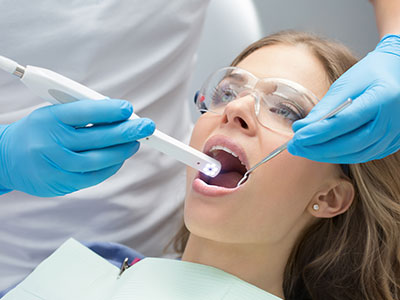
An intraoral camera is a compact, pen-sized imaging tool designed to capture clear, full‑color pictures inside the mouth. Unlike a traditional camera, it is optimized for close‑range dental imaging, producing detailed views of tooth surfaces, gum tissue, restorations, and hard‑to‑see areas such as the back molars and the lingual surfaces of teeth. Images appear on a chairside monitor in real time, giving both clinician and patient an immediate, shared view of oral health.
These cameras typically use high-resolution sensors and specialized optics to render sharp images under the unique lighting conditions of the oral cavity. Modern intraoral cameras also often incorporate features such as autofocus, LED illumination, and adjustable magnification to capture diagnostic‑quality images quickly and reliably. For dentists, that clarity translates into more confident detection of small problems that might otherwise be missed during a visual exam alone.
Beyond raw image quality, the intraoral camera is valued for its versatility. It supports routine screenings, documents progressive changes over time, and supplements other diagnostic tools like digital radiography and CBCT. When used as part of a comprehensive exam, it helps clinicians build a clearer, more complete picture of a patient’s oral health.
The intraoral camera enhances diagnostic accuracy by revealing subtle signs of disease that are difficult to catch with the unaided eye. Hairline cracks, early demineralization, marginal breakdown around fillings or crowns, and localized soft tissue changes show up with much greater definition on digital images. Catching these issues at an early stage makes it possible to address them with minimally invasive treatments and to prevent progression.
Images from the camera can be frozen, enlarged, and analyzed from multiple angles, which helps clinicians differentiate between staining and decay or between superficial wear and structural fracture. This level of detail supports more precise treatment planning—whether that means targeted restoration, monitoring, or referral to a specialist for complex concerns. The camera is especially useful for monitoring areas over time and confirming the stability of restorations and periodontal tissue.
Because intraoral cameras integrate with electronic health records, captured images become part of the patient’s permanent record. That documentation is valuable for continuity of care, allowing future providers to see baseline images and assess changes objectively. In short, the camera is both a clinical detective and a record‑keeping tool.
Seeing is understanding. For many patients, a verbal explanation of a condition can be abstract; a detailed image makes the situation tangible. Chairside photos let clinicians point out specific findings, illustrate the exact location of concern, and walk patients through proposed treatment steps while everyone is looking at the same image. This shared visual context promotes clearer conversations and helps patients make informed decisions about their care.
Using intraoral images during consultations also supports transparency. When a clinician can show a close‑up of a worn area, a fracture, or an early cavity, patients gain a better sense of urgency and the rationale for recommended care. That visual evidence reduces guesswork and aligns expectations, which typically leads to smoother treatment planning and higher patient satisfaction.
Because images are saved, clinicians can review progress with patients at follow‑up visits. Comparing current photos with prior ones makes it easier to demonstrate improvement, confirm stability, or justify a change in approach if a condition is evolving. This visual continuity fosters trust and helps patients engage more actively in preventive and restorative strategies.
Intraoral cameras are not standalone devices; they function as part of a broader digital dental ecosystem. Captured photos can be linked directly to a patient’s chart, attached to clinical notes, and shared with dental laboratories to guide restorative work. When crowns, bridges, or implant restorations are planned, high‑quality intraoral images complement digital impressions and lab specifications, helping technicians reproduce accurate shade, contour, and occlusal details.
For interdisciplinary care, images can be exported for consultation with specialists—periodontists, endodontists, or oral surgeons—streamlining the referral process and ensuring everyone involved has the same visual reference. That reduces ambiguity and improves the precision of collaborative treatment plans. In legal or administrative contexts, dated images provide objective evidence of clinical findings when documentation is required.
Practically speaking, integration reduces redundant steps and improves efficiency. Instead of describing a problem verbally or relying on sketches, clinicians can attach photographs that communicate detail instantly. This efficiency benefits patients and the clinical team by saving time and reducing room for misinterpretation.
Using an intraoral camera is quick, noninvasive, and generally comfortable. During an exam, the clinician or dental assistant will move the camera around the mouth while you watch the images on a monitor. Each snapshot takes only seconds, and most assessments require just a handful of images. The process is safe and suitable for patients of all ages, including those with special needs when handled with appropriate care and patience.
Infection control is a priority: disposable protective sleeves and barriers are used on the camera for every patient, and the outer surfaces are cleaned between appointments according to standard sterilization protocols. These precautions ensure that the device can be used routinely without increasing risk. Because the camera is a visualization tool rather than a treatment instrument, there is no direct contact that would cause tissue trauma.
After images are captured, they’re reviewed with the patient and stored securely in the dental record. This approach preserves privacy while making the photographs available for future visits or for use in treatment planning. If additional imaging is needed—such as radiographs or CBCT scans—those modalities are used in concert with intraoral photos to provide a comprehensive diagnostic picture.
Summary: Intraoral cameras give clinicians and patients a sharper, shared understanding of oral health. By revealing details that might otherwise go unnoticed, supporting clear communication, and integrating smoothly with digital workflows, this technology enhances diagnosis, treatment planning, and long‑term record keeping. If you’d like to learn more about how our office uses intraoral imaging to improve care, please contact us for more information.
Bella Vida Dental is committed to combining modern technology with patient‑centered care to help you achieve and maintain a healthy smile. Contact us to discuss how intraoral imaging may play a role in your next visit.

An intraoral camera is a compact, pen-sized digital device designed to capture high-resolution images inside the mouth for clinical evaluation. The camera uses a small sensor, specialized optics, and integrated LED illumination to produce clear, full-color photos of tooth surfaces, gum tissue, restorations, and other areas that are difficult to see with the unaided eye. Images are transmitted in real time to a chairside monitor so the clinician and patient can view the mouth together and discuss findings.
The device often includes features such as autofocus and adjustable magnification to highlight fine detail like hairline cracks, marginal breakdown, and early demineralization. Because the images are digital, they can be paused, enlarged, annotated, and stored as part of the patient record to support diagnosis and ongoing monitoring. In short, an intraoral camera extends the clinician's view and creates a shared visual record of oral health.
An intraoral camera reveals subtle signs of disease that can be missed during a visual exam, including small fractures, early decay, and localized soft tissue changes. High-resolution photos enable clinicians to magnify suspicious areas, compare different angles, and differentiate between staining and true structural damage. This level of visual detail supports earlier, more conservative interventions when appropriate.
Because images are saved and dated, clinicians can track changes over time and identify progressive issues before they become symptomatic or extensive. Early detection frequently allows for minimally invasive treatments and better long-term outcomes, reducing the likelihood of more complex procedures later. The camera therefore functions as both a diagnostic aid and a preventive tool within routine care.
No, an intraoral camera complements rather than replaces radiographic imaging such as X-rays or CBCT scans. Photographs provide detailed surface views and color information that radiographs do not, while X-rays and CBCT reveal internal structures like tooth roots, bone levels, and interproximal decay beneath the enamel. Together, these tools create a more complete diagnostic picture than either modality alone.
Clinicians integrate intraoral photos with radiographic findings to refine diagnoses and treatment plans, using each modality for its strengths. For example, a camera image can document a visible crack or margin discrepancy while an X-ray evaluates underlying tooth structure or bone. Coordinated use of both technologies improves accuracy and confidence in clinical decision-making.
An intraoral camera exam is quick, noninvasive, and usually comfortable for patients of all ages. During the assessment, the clinician or assistant will move the camera around the mouth while you watch images appear on a monitor; each photograph takes only a few seconds and most evaluations require only a handful of shots. The process does not involve radiation and causes no tissue trauma because the camera is a visualization tool rather than a treatment instrument.
After images are captured, the clinician will review them with you, pointing out findings and explaining any recommended next steps while you view the same images. Photographs can help clarify the nature and location of a problem and support shared decision-making about treatment or monitoring. If additional diagnostic imaging is needed, those tests will be discussed and coordinated with the photographic findings.
Intraoral photos are stored digitally and linked to the patient’s chart within secure practice software or an electronic health record system. Access to these images is restricted to authorized clinical staff according to privacy and security policies, and routine backups and secure storage help preserve the integrity of the files. Dated photographs become part of the permanent record and are useful for continuity of care, documentation, and clinical review.
When images are used for treatment planning or shared with other providers, the practice follows applicable privacy standards to protect patient information. Secure channels and encrypted transfers are employed for consultations or lab communication, and clinicians obtain necessary consents before using images for purposes beyond direct care. These safeguards help ensure that photographic documentation benefits clinical outcomes while maintaining confidentiality.
Yes, intraoral images are a practical tool for collaboration with specialists and dental laboratories because they provide a clear visual reference that supplements written notes and radiographs. Clinicians can attach photos to referral records or export them securely to a specialist to illustrate the exact location and appearance of a concern, which improves the precision of interdisciplinary treatment planning. For laboratory work, images help technicians match shade, contour, and occlusal relationships when fabricating restorations.
Sharing images reduces ambiguity and streamlines communication in complex cases, leading to more accurate restorations and coordinated care. When a case requires outside input, clinicians typically include dated photographs alongside radiographs and digital impressions so all providers have the same visual context. These coordinated materials support efficient, consistent outcomes across the restorative workflow.
Intraoral cameras are safe, noninvasive, and generally well tolerated by children and patients with special needs when used with appropriate technique and patience. The device is small and lightweight, and clinicians can adapt positioning and pace to accommodate comfort, limited opening, or sensory sensitivities. Disposable sleeves and other infection control measures mean the camera can be used routinely without added risk to the patient.
For very young patients or those with pronounced anxiety or behavioral challenges, clinicians may take extra time, use gentle desensitization strategies, or capture fewer images per visit to minimize stress. Photographs can be a helpful educational tool for caregivers and older children, showing concerns visually and supporting conversations about home care and follow-up. Overall, the technology is versatile and can be tailored to individual needs.
Intraoral photographs create a shared visual reference that makes clinical findings concrete and easier to understand than verbal descriptions alone. Chairside images enable clinicians to point out specific issues, demonstrate the precise location of a problem, and explain treatment options while the patient sees the same photos. This transparency helps patients make informed decisions and fosters clearer expectations about recommended care.
Saved images also allow clinicians to illustrate progress or stability at subsequent visits by comparing new photos with earlier ones. That continuity supports preventive discussions and can confirm the effectiveness of a given treatment approach. By improving clarity and documentation, intraoral imaging strengthens communication and supports collaborative planning between clinician and patient.
Infection control for intraoral cameras relies on a combination of disposable barriers, surface cleaning, and adherence to manufacturer guidelines for disinfection. Clinicians place single-use plastic sleeves or sheaths over the camera for each patient and remove them immediately after use, then clean and disinfect the camera housing and any reusable components between appointments. These procedures follow standard sterilization protocols to prevent cross-contamination while preserving device function.
Practice staff also perform routine maintenance checks on optics, illumination, and connections to ensure consistent image quality and reliable performance. Following the manufacturer's recommended cleaning agents and methods protects both patient safety and the longevity of the equipment. Regular calibration and software updates further help maintain diagnostic accuracy.
If you are interested in intraoral imaging, mention it when scheduling your visit or bring it up during your check-in so the clinical team can plan for a photo-assisted exam. Many clinicians include intraoral photos as part of a comprehensive evaluation; if you have particular concerns—such as a visible chip, stain, or sensitivity—requesting chairside photos can help clarify the issue and guide next steps. Communicating your preference ahead of time ensures the team can allocate a few extra minutes for imaging if needed.
For questions about how intraoral imaging is used in treatment planning or to learn more about the technology, contact Bella Vida Dental and ask to speak with a member of the clinical team. They can explain how photos are captured, stored, and incorporated into your record so you know what to expect during your visit.
Ready to schedule your next dental appointment or have questions about our services?
Contacting Bella Vida Dental is easy! Our friendly staff is available to assist you with scheduling appointments, answering inquiries about treatment options, and addressing any concerns you may have. Whether you prefer to give us a call, send us an email, or fill out our convenient online contact form, we're here to help. Don't wait to take the first step towards achieving the smile of your dreams – reach out to us today and discover the difference personalized dental care can make.