Oral cancer can develop quietly, often without pain in its earliest stages. That silence is what makes proactive screening so important: lesions and tissue changes spotted early are far more likely to be treated successfully. Regular visual exams remain essential, but adjunctive technologies help clinicians detect subtle abnormalities before they become advanced disease.
Screening is not a diagnostic endpoint but a critical step in a continuum of care. When suspicious findings appear, timely follow-up with biopsy, targeted imaging, or referral to a specialist can clarify the diagnosis and guide management. The goal is to catch disease at a stage where treatment is less invasive and outcomes are substantially better.
Education and routine checks together form the strongest defense. Patients who understand warning signs and attend scheduled dental exams give clinicians the best chance to identify anomalies early. This shared vigilance—between patient and dental team—improves the odds of a favorable result.
VELscope® is an adjunctive screening device that enhances the clinician’s ability to visualize changes in oral soft tissue. Using a concentrated blue light, the hand-held instrument causes healthy oral mucosa to fluoresce in predictable patterns. Areas with altered tissue architecture, dysplasia, or early malignancy often show a loss or change of that fluorescence, helping clinicians spot areas that warrant closer inspection.
Unlike diagnostic tests that require tissue removal, VELscope® is noninvasive and painless. It does not replace biopsy or pathology but serves as a practical, real-time aid during routine exams. Because it can reveal abnormalities that are not visible under normal lighting, it expands the clinician’s observational toolkit.
The technology is especially useful for monitoring patients with known risk factors or previously observed lesions. When combined with careful clinical examination and history-taking, VELscope® contributes meaningful information that supports clinical decision-making and individualized patient care.
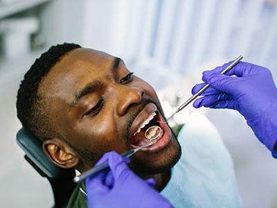
A VELscope® screening is quick and comfortable. After a standard visual inspection and palpation, your clinician dims the room lights and uses the device to illuminate the oral tissues. The bright, blue excitation light is brief and does not produce heat or discomfort; you may simply notice a stronger light and the clinician’s focused attention on specific areas.
Screenings generally take only a few minutes, and results are available immediately to the clinician. If an area shows altered fluorescence, the dentist will describe what they see, document the finding, and explain the recommended next steps—whether that means monitoring, photographing, referring, or pursuing diagnostic testing such as a biopsy.
Patients are encouraged to ask questions during the exam. Clinicians can demonstrate what’s being observed and explain why an area may appear suspicious under fluorescence. This transparency helps patients understand the reasoning behind follow-up recommendations and fosters confidence in the screening process.
An abnormal fluorescence pattern does not automatically indicate cancer; it signals tissue change that requires further evaluation. Causes for fluorescence loss include inflammation, prior trauma, infection, pigment changes, or premalignant and malignant transformations. Distinguishing among these possibilities relies on clinical skill, additional testing, and, when appropriate, pathology.
When VELscope® identifies a concern, the clinician will determine the most suitable next step. Options include close observation with short-interval rechecks, photographic documentation to track progression, targeted imaging, or referral for a biopsy. The emphasis is on prompt, evidence-based action that balances caution with the avoidance of unnecessary procedures.
Coordination of care is important when specialist input is needed. Your dental team can facilitate referrals to oral medicine specialists, head and neck surgeons, or ENT physicians for advanced diagnostics and treatment planning. Timely communication among providers ensures clarity and continuity from screening through any required treatment.
VELscope® is most effective when it complements a comprehensive prevention-focused approach. Regular dental cleanings, thorough oral examinations, and risk-factor counseling (such as tobacco and alcohol use, sun exposure for lip cancer, or HPV-related risks) create a context in which screening adds measurable value. Together, these elements support early detection and long-term oral health.
At Bella Vida Dental, clinicians integrate VELscope® into routine wellness visits for patients who would benefit from enhanced screening. This integration is part of a broader commitment to whole-patient care: assessing symptoms, reviewing medical history, and tailoring screening frequency to individual risk profiles.
Ongoing surveillance is especially important for individuals with a history of oral lesions, prior cancer, or persistent risk factors. Scheduled follow-ups and clear documentation allow clinicians to track changes over time and intervene when patterns suggest progression, maximizing the chances of early, effective treatment.
In summary, VELscope® cancer screening is a safe, noninvasive tool that enhances routine oral examinations by highlighting tissue changes that might otherwise be missed. When used thoughtfully alongside clinical judgment and appropriate follow-up, it strengthens early-detection efforts and supports better outcomes. If you’d like to learn more about VELscope® screening or how it fits into comprehensive oral care, please contact us for more information.


VELscope cancer screening is an adjunctive oral exam tool that helps clinicians visualize changes in the soft tissues of the mouth using a concentrated blue light. The device causes healthy oral mucosa to fluoresce in characteristic patterns, while areas with altered tissue architecture often show a loss or change in that fluorescence. It is designed to enhance observation during routine exams and to draw attention to areas that may warrant closer evaluation.
VELscope is not a diagnostic test and does not replace biopsy or pathology; rather, it serves as a real-time aid to the clinical examination. When the device highlights an area of concern, the clinician will use additional clinical judgment, documentation, and, if needed, further testing to determine the next steps. The goal is to increase the likelihood of identifying subtle abnormalities earlier in the continuum of care.
During a VELscope exam the clinician dims the room lights and uses the handheld device to illuminate the oral tissues with a specific blue excitation light. This light causes normal tissue to fluoresce and makes deviations from that pattern more apparent, allowing the clinician to see differences in tissue composition or structure. Observed changes are interpreted alongside the visual exam and patient history to form a more complete clinical picture.
The process is quick and incorporated into the standard oral inspection and palpation routine, so findings are available to the clinician immediately. Photographic documentation may be taken to track an area over time, and the clinician will explain any observed abnormalities and recommend appropriate follow-up. VELscope findings are one piece of information used to guide clinical decision-making rather than a stand-alone diagnosis.
VELscope screening is noninvasive and typically painless for patients. The examination involves only brief exposure to a blue light; it does not involve tissue removal, radiation, or discomfort associated with surgical procedures. Most people describe the experience as simply noticing a brighter light focused on different areas of the mouth.
Because the test is visual and noncontact, it is well suited for routine screenings and follow-up visits where clinicians need additional information without subjecting the patient to invasive testing. Should an area appear suspicious under fluorescence, the clinician may recommend further evaluation such as photographic monitoring, targeted imaging, or biopsy, which have their own processes and considerations. Clear communication during the exam helps patients understand what is being observed and why additional steps might be advised.
VELscope screening can be beneficial for a broad range of patients but is particularly useful for individuals with known risk factors for oral cancer. Examples of higher-risk patients include those with a history of tobacco or heavy alcohol use, previous oral lesions or cancer, persistent unexplained sores, significant sun exposure to the lips, or known exposure to high-risk strains of human papillomavirus (HPV). Clinicians also use the device for patients who report symptoms such as persistent pain, numbness, or lumps in the mouth or throat.
For average-risk patients, VELscope may be offered as part of a comprehensive oral exam to add an extra layer of surveillance. Ultimately, the recommendation to use VELscope depends on individual risk profile, medical history, and the clinician’s judgment. Patients are encouraged to discuss their concerns and risk factors with their dental team to determine whether adjunctive screening is appropriate.
A VELscope screening typically takes only a few minutes and is performed during a routine oral examination. The clinician will first perform a standard visual inspection and palpation, then lower the lights briefly to use the VELscope device to examine the oral tissues under the blue excitation light. Because results are immediate, the clinician can discuss any observations with the patient right away.
If an area demonstrates altered fluorescence, the clinician will explain what was seen, document the finding, and discuss recommended next steps which may include monitoring, photographic documentation, referral, or diagnostic testing. The brief nature of the test and its integration into the regular visit make it an accessible option for enhancing oral surveillance without significant additional appointment time.
An abnormal fluorescence pattern on VELscope screening indicates tissue change but is not a definitive diagnosis of cancer. Causes for loss or alteration of fluorescence can include inflammation, prior trauma, infection, benign pigment changes, premalignant changes, or malignancy. Distinguishing among these possibilities requires clinical correlation, further testing when indicated, and sometimes referral for specialist evaluation.
When an abnormality is detected the clinician will determine the most appropriate follow-up, which may involve short-interval rechecks, photographic monitoring to assess change over time, targeted imaging, or biopsy for histologic diagnosis. Prompt, measured action based on clinical judgment helps ensure that suspicious findings are evaluated thoroughly while avoiding unnecessary procedures when possible.
At Bella Vida Dental, VELscope is used as an adjunct to traditional visual examination and palpation to enhance the detection of subtle tissue changes. The device is integrated into wellness visits for patients who would benefit from enhanced surveillance, particularly those with risk factors or a history of oral lesions. It complements, rather than replaces, the clinician’s comprehensive assessment and clinical judgment.
When VELscope highlights an area of concern the team documents findings and discusses next steps tailored to the patient’s history and risk profile. If additional evaluation is warranted the practice coordinates appropriate referrals and testing to ensure timely and cohesive care, emphasizing early detection and continuity across providers when needed.
VELscope has important limitations that patients should understand: it is an adjunctive tool and not a definitive diagnostic test, and it can yield false positives or false negatives depending on the underlying tissue changes. Some benign conditions may alter fluorescence and appear suspicious, while very early or certain types of lesions might not produce a noticeable fluorescence change. Clinical context and further evaluation are essential to interpret findings accurately.
There are minimal direct risks from the VELscope exam itself since it is noncontact and noninvasive; the primary considerations involve the downstream process of following up on findings. Follow-up may require additional tests such as biopsies, which carry their own risks and implications. Clinicians balance the need for timely diagnosis with the goal of avoiding unnecessary procedures, and they communicate clearly with patients about the rationale for recommended next steps.
No special preparation is typically required for a VELscope screening beyond maintaining routine oral hygiene and arriving for your dental appointment. If you have a current sore, recent oral surgery, or acute oral infection, tell the clinician so they can interpret any findings in context and decide whether screening at that visit is appropriate. Because VELscope is performed in dimmed lighting, patients should simply be comfortable and willing to open and move the tongue and cheeks as instructed.
If you have a history of oral lesions or recent changes in your mouth, bring a concise summary of those issues and any relevant medical information to the appointment. Open communication about symptoms, tobacco or alcohol use, HPV history, and prior treatments helps the clinician tailor the exam and follow-up plan to your individual needs.
The frequency of VELscope screening depends on your individual risk factors and the clinical judgment of your dental provider; it is not a one-size-fits-all recommendation. Many patients receive adjunctive screening annually as part of their routine oral exam, while those with prior lesions, ongoing risk factors, or suspicious findings may be monitored more frequently—often at three- to six-month intervals. Your clinician will develop a surveillance schedule that balances vigilance with practicality.
If VELscope highlights a suspicious area the next steps can range from close observation and photographic monitoring to referral for targeted imaging or biopsy for histologic diagnosis. Timely follow-up and clear communication are key, and your dental team can explain the rationale for each option and coordinate referrals when specialist evaluation or treatment is recommended. For more information or to discuss screening intervals, please contact Bella Vida Dental to speak with your dental team in Tucson, AZ.

Ready to schedule your next dental appointment or have questions about our services?
Contacting Bella Vida Dental is easy! Our friendly staff is available to assist you with scheduling appointments, answering inquiries about treatment options, and addressing any concerns you may have. Whether you prefer to give us a call, send us an email, or fill out our convenient online contact form, we're here to help. Don't wait to take the first step towards achieving the smile of your dreams – reach out to us today and discover the difference personalized dental care can make.